السكري للداء مرافقة فقاعات diabeticorum= Bullosis
|
|
|
- Marylou Horton
- 5 years ago
- Views:
Transcription
1 1 / 6
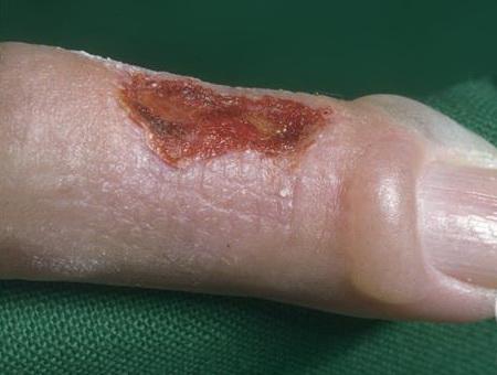
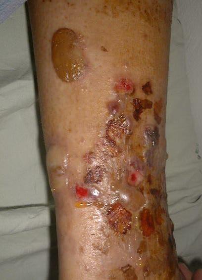
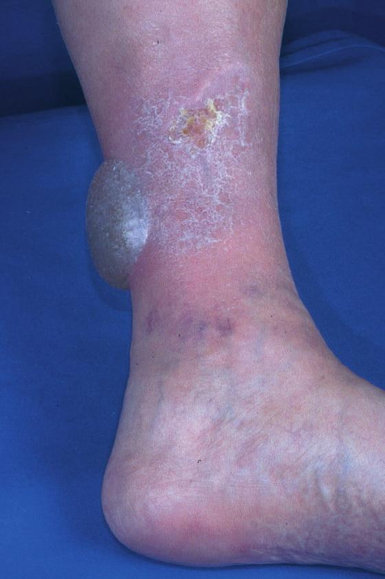
2 Bullosis diabeticorum Bullous disease of diabetes (bullosis diabeticorum) is a distinct, spontaneous, noninflammatory, blistering condition of acral skin unique to patients with diabetes mellitus. Kramer first reported bullouslike lesions in diabetic patients in ; Rocca and Pereyra first characterized this as a phlyctenar (appearing like a burn-induced blister) in Cantwell and Martz are credited with naming the condition, bullous diabeticorum, in It is also termed bullous disease of diabetes and diabetic bullae. Also see the emedicine articles Diabetes Mellitus, Type 1 and Diabetes Mellitus, Type 2. Pathophysiology The etiology of bullous disease of diabetes (bullosis diabeticorum) is not known. Patients with diabetes have been shown to have a lower threshold for suction-induced blister formation, and because of the acral prominence of diabetic bullae, the role of trauma has been speculated; however, this alone does not explain the often spontaneous development of multiple lesions at several locations. The pathophysiology is likely multifactorial. Many, but not all, patients with bullous disease of diabetes (bullosis diabeticorum) have nephropathy or neuropathy; some authors have hypothesized an etiologic association, possibly related to a local, subbasement, membrane-zone, connective-tissue alteration. Hyalinosis of small vessels noted on biopsy specimens has led some authorities to speculate microangiopathy-associated blister induction. In some, the blisters are related to UV exposure, especially in patients with nephropathy. 4 Glycemic control does not appear to have a direct correlation with blister formation. Some electron microscopic evidence has suggested an abnormality in anchoring fibrils. A reduced threshold to suction-induced blister formation in diabetic persons as compared with 2 / 6
3 nondiabetic controls has been reported. 5 Prominent acral accentuation of these lesions suggests a susceptibility to trauma-induced changes, but the definitive explanation awaits elucidation History Bullous disease of diabetes blisters occur spontaneously and abruptly, often over night, and usually without known antecedent trauma. Bullous disease of diabetes lesions tend to be asymptomatic, although mild discomfort or burning has been described. Bullous disease of diabetes blisters heal spontaneously within 2-6 weeks of onset. Physical Bullous disease of diabetes (bullosis diabeticorum) manifests as tense, nontender blisters arising on nonerythematous skin. Some blisters may be flaccid. Bullous disease of diabetes blisters typically occur on the feet or lower legs (see below), but they also may occur on fingers, toes, hands, and arms.. Causes 3 / 6
4 Prominent acral accentuation of bullous disease of diabetes lesions suggests a susceptibility to trauma-induced changes, but the definitive explanation awaits elucidation. Neuropathology, UV exposure, and microangiopathy associated with diabetes also are thought to play a role. Laboratory Studies Consider evaluation of porphyrin levels if lesions prominently involve the hands. Elevated levels may indicate porphyria cutanea tarda. Levels reportedly are normal in persons with bullous disease of diabetes (bullosis diabeticorum). If the bullous disease of diabetes blister fluid is cloudy instead of clear, consider excluding secondary bacterial infection with culture of the blister fluid. Other Tests Immunofluorescence: No primary immunologic abnormality is noted. Nonspecific capillary-associated immunoglobulin M and C3 have been reported rarely. 8 Immunofluorescenc e findings have not been consistently reproduced by others, and direct immunofluorescence findings are usually negative. 9 This study may be required to exclude clinically similar conditions (eg, bullous pemphigoid, epidermolysis bullosa acquisita) that typically show deposition of C3 and immunoglobulin G along the basement membrane zone. Procedures Skin biopsy - Consider shave biopsy or excisional/incisional biopsy to help distinguish bullous disease of diabetes (bullosis diabeticorum) from clinically similar conditions. 4 / 6
5 - For routine histologic sections, include the blister and portions of the underlying dermis in the biopsy specimen, and submit it in formalin. For biopsy findings, see Histologic Findings below. - Histologic features of bullosis diabeticorum are not entirely specific; consider direct immunofluorescence studies to exclude histologically similar entities (eg, noninflammatory bullous pemphigoid, epidermolysis bullosa acquisita, porphyria cutanea tarda). Include perilesional uninvolved skin in biopsy for direct immunofluorescence and submit it in special transport medium (eg, Michel). Histologic Findings Lesions of bullous disease of diabetes (bullosis diabeticorum) have a heterogeneous histologic presentation. The blister plane may appear in a subcorneal, intraepidermal, or subepidermal location. Histology of fresh blisters tends to show an epidermal-dermal separation (see image below). Many of the reported cases describe a separation in the superficial epidermis, within the superficial part of the spinous layer. The variable blister plane may be related to the blister age because reepithelialization can occur within days of blister onset. The blister cavity contains sterile proteinaceous fluid; an inflammatory component is absent or insignificant. Surrounding epidermis does not show significant change; however, rare reports describe associated spongiosis and degenerative keratinocytic pallor. Acantholysis is absent. Dermal changes (eg, capillary wall thickening, dermal sclerosis) may reflect the patient's underlying diabetes mellitus (see image below). Caterpillar bodies typical of porphyria have been reported in lesions of bullosis diabeticorum. 5 / 6
6 Electron microscopy of fresh blisters reveals separation in a subepidermal location, residing in the lamina lucida or the sublamina densa. 10 Anchoring fibrils and hemidesmosomes are reported absent or decreased in early blisters Medical Care Specific treatment of bullous disease of diabetes (bullosis diabeticorum) is unnecessary because the condition is self-limiting. The blister should be left intact whenever possible to serve as a sterile dressing and to avoid secondary infection. Secondary staphylococcal infections may occur, requiring antibiotic therapy. Helpful guidelines from the American Diabetes Association related to the management of diabetes are as follows: - Nutrition recommendations and interventions for diabetes: a position statement of the American Diabetes Association 11 - Standards of medical care in diabetes. I. Classification and diagnosis 12 - Standards of medical care in diabetes. V. Diabetes care. 13 Surgical Care Aspiration of fluid from bullous disease of diabetes (bullosis diabeticorum) lesions with sterile technique using a small-bore needle may be beneficial to prevent accidental rupture. Immobilization may prevent damage to the blister. Secondary tissue necrosis may necessitate debridement and possible tissue grafting. Aggressive wound healing intervention, as is enacted with diabetic ulcers, is critical, should the blister become unroofed. 6 / 6
Case Report 2 Cases of Bullosis Diabeticorum following Long-Distance JourneysbyRoad:AReportof2Cases
 Case Reports in Endocrinology Volume 2012, Article ID 367218, 5 pages doi:10.1155/2012/367218 Case Report 2 Cases of Bullosis Diabeticorum following Long-Distance JourneysbyRoad:AReportof2Cases Fatima
Bullosis diabeticorum in median nerve innervated fingers shortly after carpal tunnel release: case report.
 Bullosis diabeticorum in median nerve innervated fingers shortly after carpal tunnel release: case report. Brogren, Elisabeth; Dahlin, Lars Published in: Journal of Hand Surgery DOI: 10.1016/j.jhsa.2014.09.014
Case Rep Dermatol 2009;1:66 70 DOI: / Key Words Coma Blister Barbiturate Overdose Meningoencephalitis
 66 Coma Blisters Joana Rocha a Teresa Pereira a Filipa Ventura a Fernando Pardal b Celeste Brito a Departments of a Dermatology and b Pathology, Hospital de São Marcos, Braga, Portugal Key Words Coma Blister
DO NOT DUPLICATE. Bullosis Diabeticorum: Is There a Correlation Between Hyperglycemia and This Symptomatology? Review
 Review WOUNDS 2012;24(12):350 355 From the Barry University School of Podiatric Medicine and Surgery, Miami Shores, FL Address correspondence to: Thomas C. Wilson, BHS Barry University School of Podiatric
Dr Saleem Taibjee. Consultant Dermatologist & Dermatopathologist
 Dr Saleem Taibjee saleem.taibjee@dchft.nhs.uk Consultant Dermatologist & Dermatopathologist Case S14-10797 and S15-4023 F50. Previous blistering, now marked milia on dorsum of hands. 4mm punch biopsy The
Epidermolysis Bullosa Acquisita
 Introduction Epidermolysis Bullosa Acquisita Pages with reference to book, From 192 To 194 Nasser Rashid Dar ( Departments of Dermatology, Combined Military Hospital, Peshawar. ) Ahsan Hameed, Ashfaq Ahmad
Actinic keratosis (AK): Dr Sarma s simple guide
: Dr Sarma s simple guide") Actinic keratosis (AK): Dr Sarma s simple guide Actinic keratosis is a very common lesion that you will see in your day-to-day practice. First, let me explain the name Actinic keratosis. It means keratosis
Actinic keratosis (AK): Dr Sarma s simple guide Actinic keratosis is a very common lesion that you will see in your day-to-day practice. First, let me explain the name Actinic keratosis. It means keratosis
REPORT OF EXPERIENCES:
 REPORT OF EXPERIENCES: Non Adherent Dressings in Fragile Skin Conditions: The Use of Soft Silicone Coated Polyurethane Foam Dressing (Mepilex ) in Hereditary and Acquired Bullous Skin Diseases. Hauke Schumann
Background information of DIF
 Napa Dermatopathology Meeting 2018: Immunobullous Disease Whitney A. High, MD, JD, MEng whitney.high@ucdenver.edu Professor of Dermatology & Pathology Vice-Chairman, Dermatology Director of Dermatopathology
Autoimmune Diseases with Oral Manifestations
 Autoimmune Diseases with Oral Manifestations Martin S. Greenberg DDS, FDS RCSEd Professor Emeritus Department of Oral Medicine University of Pennsylvania Disclosure Statement I have no actual or potential
Erythema gyratumrepens-like eruption in a patient with epidermolysisbullosaacquisita associated with ulcerative colitis
 Erythema gyratumrepens-like eruption in a patient with epidermolysisbullosaacquisita associated with ulcerative colitis A. España C. Sitaru* M. Pretel L. Aguado J. Jimenez# Department of Dermatology, University
Paul K. Shitabata, M.D. Dermatopathology Institute
 Paul K. Shitabata, M.D. Dermatopathology Institute Technical Considerations Storage of slides at room temperature
Original Contribution
 Direct Immunofluorescence Test of Skin Biopsy Samples Results of 204 Cases Kabir AN, 1 Das RK, 2 Kamal M 3 Direct immunofluorescence (DIF) test of skin and renal biopsy specimens is being done on regular
Index. derm.theclinics.com. Note: Page numbers of article titles are in boldface type.
 Note: Page numbers of article titles are in boldface type. A Adhesion and migration, the diverse functions of the laminin a3 subunit, 79 87 Alopecia in epidermolysis bullosa, 165 169 Amblyopia and inherited
B. Autoimmune blistering diseases
 Go Back to the Top To Order, Visit the Purchasing Page for Details formation immediately above the basal layer. The dermal papillae, which are covered by basal cells in the single layer that is left in
Skin Integrity and Wound Care
 Skin Integrity and Wound Care By Dr. Amer Hasanien & Dr. Ali Saleh Skin Integrity and Wound Care Skin integrity: the presence of normal Skin & Uninterrupted skin layers by wounds. Factors affecting appearance
Pemphigus in younger age group in Bangladeshi population
 ORIGINAL ARTICLE in younger age group in Bangladeshi population Abdul Wahab 1, MD, Lubna Khondker 1, MD, Jamal Uddin 1, MD, Ishrat Bhuiyan 2, MD Shirajul Islam Khan 3, MD, Zafrul Islam 1, MD, Rahmat Ali
Autoimmune bullous dermatoses
 Autoimmune bullous dermatoses Overview of serological diagnostics in autoimmune blister-forming diseases of the skin Pemphigoid diseases Pemphigus diseases Epidermolysis bullosa acquisita Dermatitis herpetiformis
Clinicopathological correlation of blistering diseases of skin
 Bangladesh Med Res Counc Bull 2008; 34: 48-53 Copyright 2008 by Bangladesh Medical Research Council Clinicopathological correlation of blistering diseases of skin A.K.M. Nurul Kabir 1, Mohammed Kamal 1
BSD SELF-ASSESSMENT CASES 21-24
 BSD SELF-ASSESSMENT CASES 21-24 EDINBURGH, 2 JULY 2018 LASZLO IGALI CASE 21 CLINICAL HISTORY Female, 43 years, nodule on left side of face/jaw angle area. Slow growth over years. MACRO (NOT GIVEN) Skin
HEMORRHAGIC BULLOUS HENOCH- SCHONLEIN PURPURA: A CASE REPORT
 HEMORRHAGIC BULLOUS HENOCH- SCHONLEIN PURPURA: A CASE REPORT Nirmala Ponnuthurai, Sabeera Begum, Lee Bang Rom Paediatric Dermatology Unit, Institute of Paediatric, Hospital Kuala Lumpur, Malaysia Abstract
Studies in Cutaneous Aging: II. The Microvasculature
 0022-202X/82/7805-0444$02.00/0 THE JOURNAL OF lnvestigativ E DERMATOLOGY, 78:444-448, 1982 Copyright ]982 by The Williams & Wilkins Co. Vol. 78, No.5 Printed in U.S.A. Studies in Cutaneous Aging: II. The
Saturday, 09 October :14 - Last Updated Sunday, 13 February :43
 1/9 2/9 Diabetic finding had 50.Diabetic pretibial asymmetrical superficial extravasated hyperpigmentation. behind diabetic inareas dermopathy diabetes. blood dermopathy dermopathy, erythrocytes distribution.
A case of bullous pemphigoid following pemphigus foliaceus
 #2228 A case of bullous pemphigoid following pemphigus foliaceus Priyanka Vedak MD 1, Danielle Levine MD 1,3, Lyn Duncan MD 2,3, Hensin Tsao 1,3, Daniela Kroshinsky MD MPH 1,3 1. Department of Dermatology,
CLINCOPATHOLOGICAL CASE
 CLINCOPATHOLOGICAL CASE Generalized vesiculo-bullous and pustular eruption in an adult man Hassab El-Naby H, MD, El-Khalawany M, MD Department of Dermatology, Al-Azhar University, Cairo, Egypt CLINICAL
Classification: 1. Infective: 2. Traumatic: 3. Idiopathic: Recurrent Aphthous Stomatitis (RAS) 4. Associated with systemic disease:
 4. Associated with systemic disease:") Classification: 1. Infective: 2. Traumatic: 3. Idiopathic: Recurrent Aphthous Stomatitis (RAS) 4. Associated with systemic disease: Hematological GIT Behcet s HIV 5. Associated with dermatological diseases:
Classification: 1. Infective: 2. Traumatic: 3. Idiopathic: Recurrent Aphthous Stomatitis (RAS) 4. Associated with systemic disease: Hematological GIT Behcet s HIV 5. Associated with dermatological diseases:
Acquired and Inherited Bullous Diseases
 Acquired and Inherited Bullous Diseases Erin Wei MD Brigham and Women s Hospital, Department of Dermatology Instructor, Harvard Medical School Director, Bullous Disease Clinic No disclosures Conflict of
Mr Zachary Moaveni Plastic Surgeon, Middlemore Hospital. Mr Adam Bialostocki Plastic Surgeon, Tauranga
 Mr Zachary Moaveni Plastic Surgeon, Middlemore Hospital Mr Adam Bialostocki Plastic Surgeon, Tauranga Mr. Adam Bialostocki Plastic Surgeon Minor Burns First Aid Remove the burning agent / wet clothes
STUDIES ON THE PATHOGENESIS OF EPIDERMOLYSIS BULLOSA*
 STUDIES ON THE PATHOGENESIS OF EPIDERMOLYSIS BULLOSA* ROGER W. PEARSON, M.D. Clinical classification of the diseases in the epidermolysis bullosa group has led to little progress toward elucidation of
Comparative microanatomy of the normal skin with that of immunobullous condition
 Original article: Comparative microanatomy of the normal skin with that of immunobullous condition 1Dr BananiKundu, 2 DrAnirban Sadhu, 3 DrRudradev Meyur, 4 Dr SauravKundu, 5 Dr Alpana De, 6Dr SatabdiSarkar
Immunofluorescence in Oral Dermatological Disorders- No Shiny Matter
 Journal of Academy of Dental Education Journal of Academy of Dental Education, 24-28, DOI: 10.18311/jade/2015-2016/15951 ISSN (Print): 2348-1595 ISSN (Online) : 2348-2621 Immunofluorescence in Oral Dermatological
Mosaad Megahed Histopathology of Blistering Diseases With Clinical, Electron Microscopic, Immunological and Molecular Biological Correlations
 Mosaad Megahed Histopathology of Blistering Diseases With Clinical, Electron Microscopic, Immunological and Molecular Biological Correlations Springer-Verlag Berlin Heidelberg GmbH Mosaad Megahed Histopathology
Blistering disorders and their differential diagnosis and management
 and their differential diagnosis and management STEVE KOSSARD, MB BS, PhD, FACD The type and pattern of cutaneous blisters may provide an essential clue to the diagnosis of specific dermatological disorders.
Wound Classification. Overview
 Overview Jeffrey A. Niezgoda, MD FACHM, MAPWCA, CHWS Review of Initial Wound Care Consultation Rational for Classification Wound Appearance Wound Etiology Management Algorithms Initial Wound Care Consult
Current concepts of autoimmune bullous diseases Advances in pathogenesis. Luca Borradori
 Current concepts of autoimmune bullous diseases Advances in pathogenesis Luca Borradori Dept. of Dermatology Inselspital, University Hospital of Berne Switzerland Luca.Borradori@insel.ch Autoimmune bullous
Bullous Pemphigoid with Lymphocytic Colitis: A Case Report and Short Literature Review
 Dermatol Ther (Heidelb) (2016) 6:437 441 DOI 10.1007/s13555-016-0135-4 CASE REPORT Bullous Pemphigoid with Lymphocytic Colitis: A Case Report and Short Literature Review Alexandra Sperl. Johann W. Bauer.
Recent Advances in the Molecular Pathology of Bullous Skin Disorders
 1 Bahrain Medical Bulletin, Vol. 27, No. 2, June 2005 Recent Advances in the Molecular Pathology of Bullous Skin Disorders John A McGrath* Maintenance of an intact epidermis depends on secure adhesion
Thermal Dermal Burn Modeling in Rats and Minipigs
 Thermal Dermal Burn Modeling in Rats and Minipigs Comparative Biosciences, Inc. 786 Lucerne Drive Sunnyvale, CA 94085 Telephone: 408.738.9261 www.compbio.com Premier Preclinical Contract Research Organization
DERMATOLOGICAL EMERGENCIES. DR. Ian Hoyle MBBS DIP IMC RCS (Ed), DA (UK),FRACGP,FACRRM,DIP DERM(Wales) TASMANIAN SKIN AND BODY CENTRE
, DA (UK),FRACGP,FACRRM,DIP DERM(Wales) TASMANIAN SKIN AND BODY CENTRE") DERMATOLOGICAL EMERGENCIES DR. Ian Hoyle MBBS DIP IMC RCS (Ed), DA (UK),FRACGP,FACRRM,DIP DERM(Wales) TASMANIAN SKIN AND BODY CENTRE Dermatological Emergencies INFECTIONS ERYTHRODERMA DRUG ERUPTIONS STEVENS-JOHNSON
DERMATOLOGICAL EMERGENCIES DR. Ian Hoyle MBBS DIP IMC RCS (Ed), DA (UK),FRACGP,FACRRM,DIP DERM(Wales) TASMANIAN SKIN AND BODY CENTRE Dermatological Emergencies INFECTIONS ERYTHRODERMA DRUG ERUPTIONS STEVENS-JOHNSON
Surgical Care at the District Hospital. EMERGENCY & ESSENTIAL SURGICAL CARE
 Surgical Care at the District Hospital 1 5 Basic Surgical Procedures Key Points 2 5.1 Wound Management Many important procedures can be performed under local anesthesia and do not require a surgical specialist
Histopathology: skin pathology
 Histopathology: skin pathology These presentations are to help you identify, and to test yourself on identifying, basic histopathological features. They do not contain the additional factual information
Skin lesions & Abrasions
 Skin lesions & Abrasions What Are Skin Lesions? A skin lesion is a part of the skin that has an abnormal growth or appearance compared to the skin around it Types of Skin Lesions Two types of skin lesions
EPIDERMOLYSIS BULLOSA
 EPIDERMOLYSIS BULLOSA Definition Epidermolysis bullosa (EB) is a term used to describe a group of rare mainly hereditary, chronic, non-inflammatory diseases of skin and mucous membranes. EB is characterized
Not All That Blisters Is a Burn! Jamie Hoffman-Rosenfeld, MD CHAMP Webinar December 6, 2012
 Not All That Blisters Is a Burn! Jamie Hoffman-Rosenfeld, MD CHAMP Webinar December 6, 2012 Objectives To review the epidemiology of burns in children including burns caused by abuse To review the steps
A cross-sectional study of clinical, histopathological and direct immmunofluorescence diagnosis in autoimmune bullous diseases
 Original Article A cross-sectional study of clinical, histopathological and direct immmunofluorescence diagnosis in autoimmune bullous diseases Anchal Jindal, MD 1 Rushikesh Shah, MBBS 2 Neela Patel, MD
Pharmacologyonline 1: 1-6 (2010) Case Report Ravishankar and Hiremath CIPROFLOXACIN INDUCED BULLOUS PEMPHIGOID: A CASE REPORT
 Case Report Ravishankar and Hiremath CIPROFLOXACIN INDUCED BULLOUS PEMPHIGOID: A CASE REPORT") CIPROFLOXACIN INDUCED BULLOUS PEMPHIGOID: A CASE REPORT Ravishankar AC 1*, Hiremath SV 1 1 Dept of Pharmacology and Pharmacotherapeutics, JN Medical College, Belgaum, India. Summary Bullous pemphigoid
CIPROFLOXACIN INDUCED BULLOUS PEMPHIGOID: A CASE REPORT Ravishankar AC 1*, Hiremath SV 1 1 Dept of Pharmacology and Pharmacotherapeutics, JN Medical College, Belgaum, India. Summary Bullous pemphigoid
Autoimmune bullous disorders 1)
") Clin Chem Lab Med 2006;44(2):144 149 2006 by Walter de Gruyter Berlin New York. DOI 10.1515/CCLM.2006.027 2006/39 Review Autoimmune bullous disorders 1) Rüdiger Eming* and Michael Hertl for the members
Clin Chem Lab Med 2006;44(2):144 149 2006 by Walter de Gruyter Berlin New York. DOI 10.1515/CCLM.2006.027 2006/39 Review Autoimmune bullous disorders 1) Rüdiger Eming* and Michael Hertl for the members
Journal of Pediatric Sciences
 Journal of Pediatric Sciences Severe generalized recessive dystrophic epidermolysis bullosa (Hallopeau-Siemen s) : a case report Iffat Hassan, Mohammad Abid Keen Journal of Pediatric Sciences 2013;5:e190
Abscess. A abscess is a localized collection of pus in the skin and may occur on any skin surface and be formed in any part of body.
 Abscess A abscess is a localized collection of pus in the skin and may occur on any skin surface and be formed in any part of body. Ethyology Bacteria causing cutaneous abscesses are typically indigenous
Glomerular pathology in systemic disease
 Glomerular pathology in systemic disease Lecture outline Lupus nephritis Diabetic nephropathy Glomerulonephritis Associated with Bacterial Endocarditis and Other Systemic Infections Henoch-Schonlein Purpura
and Isolation of Antibody in Linear Immunoglobulin A Bullous Dermatosis
 Identification of the Cutaneous Basement Membrane Zone Antigen and Isolation of Antibody in Linear Immunoglobulin A Bullous Dermatosis John J. Zone, Ted B. Taylor, Donald P. Kadunce, and Laurence J. Meyer
University of Groningen. Acantholysis in pemphigus van der Wier, Gerda
 University of Groningen Acantholysis in pemphigus van der Wier, Gerda IMPORTANT NOTE: You are advised to consult the publisher's version (publisher's PDF) if you wish to cite from it. Please check the
To Correlate Clinical Diagnosis with Histopathology and DIF Pattern of Autoimmune Based Vesiculobullous Disorders In A Tertiary Teaching Hospital
 IOSR Journal of Dental and Medical Sciences (IOSR-JDMS) e-issn: 2279-0853, p-issn: 2279-0861.Volume 17, Issue 7 Ver. 2 (July. 2018), PP 01-06 www.iosrjournals.org To Correlate Clinical Diagnosis with Histopathology
Subtle Signs of Child Abuse Child s Protection Office MOH Presented by Dr.Fatoumah Alabdulrazzaq M.D,FRCPC,FAAP,PEM(C)
") Subtle Signs of Child Abuse Child s Protection Office MOH Presented by Dr.Fatoumah Alabdulrazzaq M.D,FRCPC,FAAP,PEM(C) Cutaneous Injuries Bruise : injury to soft tissues in which skin is not broken, characterized
Subtle Signs of Child Abuse Child s Protection Office MOH Presented by Dr.Fatoumah Alabdulrazzaq M.D,FRCPC,FAAP,PEM(C) Cutaneous Injuries Bruise : injury to soft tissues in which skin is not broken, characterized
Crescentic Glomerulonephritis (RPGN)
") Crescentic Glomerulonephritis (RPGN) Background Rapidly progressive glomerulonephritis (RPGN) is defined as any glomerular disease characterized by extensive crescents (usually >50%) as the principal histologic
Crescentic Glomerulonephritis (RPGN) Background Rapidly progressive glomerulonephritis (RPGN) is defined as any glomerular disease characterized by extensive crescents (usually >50%) as the principal histologic
DERMATOLOGY SKIN DISEASE: APPROACH TO DIAGNOSIS
 DERMATOLOGY SKIN DISEASE: APPROACH TO DIAGNOSIS History Clinical Examination List and Prioritise Differentials Diagnostic Testing/Trials (eg Treatment Trial) Correlate All Findings History Signalment age,
건강한성인에서의오진하기쉬운포도구균성열상피부증후군의치험례. Staphylococcal Scalded Skin Syndrome in a Healthy Adult: Easy to Misdiagnose
 Archives of Hand and Microsurgery Arch Hand Microsurg 2018;23(4):271-276. https://doi.org/10.12790/ahm.2018.23.4.271 pissn 2586-3290 eissn 2586-3533 Case Report 건강한성인에서의오진하기쉬운포도구균성열상피부증후군의치험례 김홍일ㆍ곽찬이ㆍ박언주
Diabetic Foot Ulcers. Alex Khan APRN ACNS-BC MSN CWCN CFCN WCN-C. Advanced Practice Nurse / Adult Clinical Nurse Specialist
 Diabetic Foot Ulcers Alex Khan APRN ACNS-BC MSN CWCN CFCN WCN-C Advanced Practice Nurse / Adult Clinical Nurse Specialist Organization of Wound Care Nurses www.woundcarenurses.org Objectives Identify Diabetic/Neuropathic
BULLOUS SYSTEMIC lupus erythematosus
 OBSERVATION Bullous Systemic Lupus Erythematosus With Autoantibodies Recognizing Multiple Skin Basement Membrane Components, Bullous Pemphigoid Antigen 1, Laminin-5, Laminin-6, and Type VII Collagen Lawrence
B. Incorrect! The ectoderm does not produce the dermis. C. Incorrect! The dermis is derived from the mesoderm.
 Human Anatomy - Problem Drill 04: The Integumentary System Question No. 1 of 10 Instructions: (1) Read the problem and answer choices carefully, (2) Work the problems on paper as 1. From the inner cell
Skin Deep. Agenda. Burns Wounds Debridement Evaluation and Management Services. Presented by: Mike Strong, SFM The Work Comp Experts.
 Presented by: Mike Strong, SFM The Work Comp Experts Agenda Wounds Debridement Evaluation and Management Services 2 1 Types of First Degree Second Degree Third Degree Rule of 9 Adults Infants Burn Coding
Hemostasis Inflammatory Phase Proliferative/rebuilding Phase Maturation Phase
 The presenters are staff members of the CHI Health St. Elizabeth Burn and Wound Center. Many of the products discussed are used in our current practice but we have no conflict of interest to disclose.
2 Anonychia/Micronychia
 2 Anonychia/Micronychia Total or partial absence of the nail May be congenital or acquired Table 2.1. Causes of anonychia/micronychia Congenital Acquired Trauma Amniotic bands Bullous Teratogens (drugs,
Indian Journal of Dermatopathology and Diagnostic Dermatology
 Volume 1 Issue 1 Jan-Jun 2014 Online full text at www.ijdpdd.com Indian Journal of Dermatopathology and Diagnostic Dermatology DSI IADVL Karnataka Branch Official Publication of Dermatopathology Society
Cutaneous Conditions Associated with Systemic Disease
 Cutaneous Conditions Associated with Systemic Disease Johnnie M Woodson, M.D., F.A.A.D. Assistant Professor of Dermatology University of Nevada School of Medicine Director of J. Woodson Dermatology & Associates,
Skin and Body Membranes
 Essentials of Human Anatomy & Physiology Elaine N. Marieb Seventh Edition Chapter 4 Skin and Body Membranes Slides 4.1 4.32 Lecture Slides in PowerPoint by Jerry L. Cook Skin and Body Membranes Function
Pathology of the skin. 2nd Department of Pathology, Semmelweis University
 Pathology of the skin 2nd Department of Pathology, Semmelweis University Histology of the skin Epidermis: Stratum corneum Stratum granulosum Stratum spinosum Stratum basale Dermis: papillary and reticular
Determining Wound Diagnosis and Documentation Tips Job Aid
 Determining Wound Diagnosis and Job Aid 1 Coding Is this a traumatic injury from an accident? 800 Codes - Injury Section of the Coding Manual Code by specific site of injury. Only use for accidents or
4 Skin and Body Membranes Study Guide
 Name: SKIN AND BODY MEMBRANES: 4 Skin and Body Membranes Study Guide Period: Body membranes, which cover body surfaces, line its cavities, and form protective sheets around organs, fall into two major
Conflicts. Objectives. University of Texas Health Science Center at San Antonio. Pediatrics Grand Rounds 24 August Pediatric Dermatology 101
 Pediatric Dermatology 101 John C. Browning, MD, FAAD, FAAP Conflicts Investigator: ViroXis Advisor: ViroXis Advisory Board: TopMD Speaker: Galderma Objectives Understand the meaning and importance of cutaneous
Immunobullous Diseases: Review and Update. May P. Chan, MD Associate Professor of Pathology and Dermatology University of Michigan
 Immunobullous Diseases: Review and Update May P. Chan, MD Associate Professor of Pathology and Dermatology University of Michigan Diagnosis of Immunobullous Diseases Clinical H&E DIF DIAGNOSIS IIF ELISA
Dóra Ujvárosy MD. Medical University of Debrecen Oxyology and Emergency Department
 Dóra Ujvárosy MD. Medical University of Debrecen Oxyology and Emergency Department Functions Definition A burn is a type of injury to the skin caused by heat, electricity, chemicals, light, radiation or
RENAL HISTOPATHOLOGY
 RENAL HISTOPATHOLOGY Peter McCue, M.D. Department of Pathology, Anatomy & Cell Biology Sidney Kimmel Medical College There are no conflicts of interest. 1 Goals and Objectives! Goals Provide introduction
MUCOCUTANEOUS LESIONS Normal structures in epithelium cell adhesion to each other and to underlying connective tissue:
 ORAL DERMATOSES AND MUCOSAL/GINGIVAL LESIONS MUCOCUTANEOUS LESIONS Normal structures in epithelium cell adhesion to each other and to underlying connective tissue: Diagram taken from: Oral and Maxillofacial
Basics of Skin Biopsy Techniques
 Basics of Skin Biopsy Techniques Kelly Quinn, D.O. Lehigh Valley Health Network/PCOM Department of Dermatology Osteopathic Medical Conference and Exposition October 7, 2017 None to disclose Conflicts of
Principles of Anatomy and Physiology
 Principles of Anatomy and Physiology 14 th Edition CHAPTER 5 The Integumentary System Introduction The organs of the integumentary system include the skin and its accessory structures including hair, nails,
LESIONS OF THE ORAL CAVITY ORAL CAVITY. Oral Cavity Subsites 4/10/2013 LIPS TEETH GINGIVA ORAL MUCOUS MEMBRANES PALATE TONGUE ORAL LYMPHOID TISSUES
 LESIONS OF THE ORAL CAVITY David I. Kutler, MD, FACS Associate Professor Division of Head and Neck Surgery Department of Otolaryngology HNS Weill Cornell Medical Center ORAL CAVITY LIPS TEETH GINGIVA ORAL
1/3/2008. Karen Burke Priscilla LeMone Elaine Mohn-Brown. Medical-Surgical Nursing Care, 2e Karen Burke, Priscilla LeMone, and Elaine Mohn-Brown
 Medical-Surgical Nursing Care Second Edition Karen Burke Priscilla LeMone Elaine Mohn-Brown Chapter 46 Caring for Clients with Burns Types of Burns Thermal Dry heat flame Moist heat steam or hot liquid
SWISS SOCIETY OF NEONATOLOGY. Neonatal blistering - a butterfly child
 SWISS SOCIETY OF NEONATOLOGY Neonatal blistering - a butterfly child August 2010 2 Kaelin S, Weibel L, Arlettaz Mieth R, Neonatal Intensive Care Unit (KS, AMR), Department of Dermatology (WL), University
Histologic Comparison of Pressure and Autoimmune Wounds
 Histologic Comparison of Pressure and Autoimmune Wounds Item Type Thesis Authors Nanda, Alisha Publisher The University of Arizona. Rights Copyright is held by the author. Digital access to this material
Skin and Body Membranes Body Membranes Function of body membranes Cover body surfaces Line body cavities Form protective sheets around organs
 Skin and Body Membranes Body Membranes Function of body membranes Cover body surfaces Line body cavities Form protective sheets around organs Classification of Body Membranes Epithelial membranes Cutaneous
Department of Dermatology, Nippon Medical School, 1-1-5, Sendagi, Bunkyo-ku, Tokyo , Japan 2
 Dermatology Research and Practice Volume 2010, Article ID 931340, 5 pages doi:10.1155/2010/931340 Case Report Paraneoplastic Pemphigus Presenting as Mild Cutaneous Features of Pemphigus Foliaceus and Lichenoid
High Risk Podiatry in a Vascular Setting; A new paradigm in Diabetic Foot Disease? Ereena Torpey Senior Podiatrist - FMC
 High Risk Podiatry in a Vascular Setting; A new paradigm in Diabetic Foot Disease? Ereena Torpey Senior Podiatrist - FMC A new paradigm? Foot ulceration 101 Assessing Perfusion a new challenge Pressure
Review Article Clinical Relevance of Autoantibodies in Patients with Autoimmune Bullous Dermatosis
 Clinical and Developmental Immunology Volume 2012, Article ID 369546, 9 pages doi:10.1155/2012/369546 Review Article Clinical Relevance of Autoantibodies in Patients with Autoimmune Bullous Dermatosis
Skin and Body Membranes
 4 Skin and Body Membranes PowerPoint Lecture Slide Presentation by Jerry L. Cook, Sam Houston University ESSENTIALS OF HUMAN ANATOMY & PHYSIOLOGY EIGHTH EDITION ELAINE N. MARIEB Skin and Body Membranes
Junctional adhesion molecule overexpression in Kaposi varicelliform eruption skin lesions - as a possible herpes virus entry site
 Case Report OPEN ACCESS Junctional adhesion molecule overexpression in Kaposi varicelliform eruption skin lesions - as a possible herpes virus entry site Ana Maria Abreu-Velez 1, MD., PhD, A. Deo Klein
COPYRIGHTED MATERIAL. Introduction CHAPTER 1. Introduction
 CHAPTER 1 Introduction OVERVIEW The clinical features of skin lesions are related to the underlying pathological processes. Broadly skin conditions fall into three clinical groups: (a) those with a well-defined
Hole s Human Anatomy and Physiology Eleventh Edition. Mrs. Hummer. Chapter 6
 Hole s Human Anatomy and Physiology Eleventh Edition Mrs. Hummer Chapter 6 1 Chapter 6 Skin and the Integumentary System Composed of several tissues Maintains homeostasis Protective covering Retards water
Acute and Chronic WOUND ASSESSMENT. Wound Assessment OBJECTIVES ITEMS TO CONSIDER
 WOUND ASSESSMENT Acute and Chronic OBJECTIVES Discuss classification systems and testing methods for pressure ulcers, venous, arterial and diabetic wounds List at least five items to be assessed and documented
Some skin conditions
 Some skin conditions Some skin conditions Acute Inflammatory Dermatoses Chronic Inflammatory Dermatoses Blistering (Bullous) Diseases Panniculitis Disorders of Epidermal Appendages -Urticaria -Acute eczematous
Burns. A Comprehensive Review Assessment & Management
 Burns A Comprehensive Review Assessment & Management 1 Objectives Understand types of Burns Understand the pathophysiology of the Burns Understand Rule of Nine Understand Classification of Burns Identify
DERMATOLOGY VOLUME 40 NUMBER 5 PART 1 MAY 1999
 CONTINUING MEDICAL EDUCATION The new pemphigus variants Journal of the American Academy of DERMATOLOGY VOLUME 40 NUMBER 5 PART 1 MAY 1999 Neha D. Robinson, MD, a Takashi Hashimoto, MD, b Masayuki Amagai,
VASCULAR WOUNDS PATHOPHYSIOLOGY AND MANAGEMENT
 VASCULAR WOUNDS PATHOPHYSIOLOGY AND MANAGEMENT Lucy Stopher, A/CNS Vascular Surgery ...it is best to think of a wound not as a disease, but rather as a manifestation of disease. Joe McCulloch In order
DMEPOS: hospital beds, bed accessories, and pressurereducing
 ACTION: Final DATE: 07/02/2018 10:03 AM 5160-10-18 DMEPOS: hospital beds, bed accessories, and pressurereducing support surfaces. (A) Definitions and explanations. (1) "Group 1," "group 2," and "group
Pimples and Boils!! Dr Nathan Harvey Anatomical Pathology, PathWest
 Pimples and Boils!! Dr Nathan Harvey Anatomical Pathology, PathWest Overview & Learning Objectives Review the cardinal signs/symptoms of acute inflammation Review the histological features of acute inflammation
Original Article ABSTRACT
 Original Article doi: 10.5146/tjpath.2015.01345 Utility of Direct Immunofluorescence Studies in Subclassification of Autoimmune Sub-Epidermal Bullous Diseases: A 2-Year Study in a Tertiary Care Hospital
ELECTRON MICROSCOPE STUDIES OF DERMAL-EPIDERMAL SEPARATION IN HUMAN SKIN*
 ELECTRON MICROSCOPE STUDIES OF DERMAL-EPIDERMAL SEPARATION IN HUMAN SKIN* ROGER W. PEARSON, M.D.t AND BENJAMIN SPARGO, M.D.t Electron microscope studies by several workers (1, 2, 3) have demonstrated an
Topic 4: Fractures and External Fixation
 Topic 4: Fractures and External Fixation Acute Compartment Syndrome Prof. Dr. Andreas Platz Stadtspital Triemli, Zürich Demographics Incidence: Men 7.3/100,000 Women 0.7/100,000 69% due to trauma 36% fx
Surgical Pathology Report
 Louisiana State University Health Sciences Center Department of Pathology Shreveport, Louisiana Accession #: Collected: Received: Reported: 6/1/2012 09:18 6/2/2012 09:02 6/2/2012 Patient Name: Med. Rec.